Amir Siahpoosh1,2 ![]() ,
Inas Soleimani2
,
Inas Soleimani2
For correspondence:- Amir Siahpoosh Email: Siahpoosh-a@ajums.ac.ir Tel:+9861333738381
Received: 11 March 2016 Accepted: 16 July 2016 Published: 30 August 2016
Citation: Siahpoosh A, Soleimani I. In vitro evaluation of inhibitory effect of Phoenix dactylifera bark extract on rat lipid peroxidation and blood hemolysis. Trop J Pharm Res 2016; 15(8):1707-1713 doi: 10.4314/tjpr.v15i8.16
© 2016 The authors.
This is an Open Access article that uses a funding model which does not charge readers or their institutions for access and distributed under the terms of the Creative Commons Attribution License (http://creativecommons.org/licenses/by/4.0) and the Budapest Open Access Initiative (http://www.budapestopenaccessinitiative.org/read), which permit unrestricted use, distribution, and reproduction in any medium, provided the original work is properly credited..
Purpose: To determine the polyphenolic content of P. dactylifera L. (date palm) bark extract and to investigate its in vitro inhibitory effects on lipid peroxidation in the brain, liver, and kidney tissues of rat, as well as on blood hemolysis in rats.
Methods: P. dactylifera L. barks were collected from Ahvaz, (Khuzestan, Iran). Methanolic extract of the P. dactylifera barks were prepared using maceration method. Total phenolic, flavonoid and proanthocyanidin contents were determined by colorimetric methods. The effects of the extract were investigated on thiobarbituric acid reactive substances formation in Fe2+/ascorbate induced-lipid peroxidation in brain, liver and kidney tissues of rats. Furthermore, the inhibitory effects of the extracts on erythrocyte hemolysis caused by 2,2'-Azobis (2-amidinopropane) dihydrochloride (AAPH) were evaluated.
Results: Total phenolic, flavonoids and proanthocyanidins contents per each g dry extract of P. dactylifera L. were 50.7 mg tannic acid, 10.38 mg rutin, and 5.45 mg cyanidin, respectively, while half-maximal inhibitory concentration (IC50) for inhibition of lipid peroxidation in brain, liver, and kidney tissues of rats was 3150.71, 1941.45, and 1546.01 µg/mL, respectively, and for the inhibition of erythrocyte hemolysis, it was 472.41 µg/mL.
Conclusion: The findings of this study indicate significant anti-lipid peroxidation and anti-hemolytic effects of the bark extract. Therefore, the extract can potentially be used for the in vivo treatment of diseases associated with lipid peroxidation such as cancers and Alzheimer's disease, but further studies are required.
Introduction
Free radicals are highly active molecules generated from reactive oxygen species (ROSs), which are produced in different conditions such as exposures to sunlight, ultraviolet ray, and from metabolic processes [1,2]. Free radicals have significant pathogenic effects such as DNA destruction, ischemic, aging, and digestive problems. Antioxidants play an important role on the health of body and through different mechanisms prevent the poisonous effects of free radicals [2].
Free radicals damage the carbohydrates, proteins, lipids, and DNA of the cell. Compared with other macromolecules, lipids are more rapidly affected by free radicals [3]. Free radicals increase the oxidation of unsaturated fatty acids at cell membrane structure, which is called lipid peroxidation and this reaction continues as a chain [4]. P. dactylifera L. (date palm) belongs the family Arecaceae. Date fruit is the most used part of the plant containing of carotenoids, tannin, phenolic compounds, flavonoids, and leucoanthocyanidin. In addition, it contains 50% sugar, sucrose, modified sugar, pyridine derivatives. Date palm grows in tropical and subtropical regions. Its fruit is used as the main food in central Asia and North Africa.
Studies have reported the antioxidant and antimutagenic properties of date fruit [5]. Although several studies have investigated antioxidant activities of date fruits, none of them investigated the antioxidant activities of the date bark. Therefore, the present study aimed to investigate the amount of phenolic compounds, anti-lipid peroxidation, and anti-hemolytic activity of Phoenix dactylifera bark.
Methods
Chemical material
The L-ascorbic acid, 2,2-azobisdihydrochloride, thiobarbituric acid (TBA), and phosphate buffered saline (PBS) were purchased from Sigma (St. Louis, USA,). The FeSO4 Anhydrous and trichloroacetic acid (TCA) were purchased from Fluka Co. (Buchs, Switzerland). All other chemicals used in this study were of analytical grade supplied by Merck (Darmstadt, Germany).
Plant materials
Fresh barks of P. dactylifera L. were collected fresh from Ahvaz (Khuzestan, Iran). The extracts were prepared according to the instructions of Iranian herbal pharmacopeia explained in the previous work [6].
Briefly, the powders were macerated in methanol for 48 h at 25-30 °C. The extracts were centrifuged at 4000 rpm for 20 min to obtain the supernatant and the remaining sample was also re-extracted under the same conditions. The combined extracts were filtered, concentrated in a rotary evaporator, and dried with a freeze dryer.
Total phenolic content determination
The total phenolic content was estimated by the Folin-Ciocalteu method as follows: 0.5 mL of extract and 2.5 mL of a 1/10 aqueous dilution of Folin-Ciocalteu reagent were mixed to form mixed copound. Following 5 min, 2 mL of 7.5 % Na2CO3 was added to the mixed compound and then incubated at room temperature for 120 min. Absorption at 765 nm was measured by using a spectrophotometer (UV-1800, Shimadzu). Total phenolic contens were determined as milligrams of tannic acid equivalents per gram of extract (mg tannic acid /g dry extract) using a standard calibration curve ranging 0 to 100 µg/ml [6].
Total flavonoid content determination
The flavonoid content was measured by the AlCl3 method as follows: The sample solution (1 mL) was added to 1 mL of 2 % methanolic AlCl3. 6H2O. The absorbance was measured 10 min later at 430 nm. The total flavonoid content was determined using a standard curve with 0 - 80 µg/ml rutin, then expressed as mg of rutin equivalents (mg rutin/g dry extract) [6].
Oligomeric proanthocyanidin content determination
To 0.5 mL sample were added 6 mL of n-butanol:HCl (95:5;v:v) and 0.2 mL of 2 % (w:v) solution of NH4Fe(SO4)2.12H2O in 2 M HCl. The tightly capped tubes were heated during 40 min at 95 ± 2 °C in a water bath. After cooling, the absorbance of colored solutions was measured at 550 nm. The results were expressed against standard cyanidin (mg cyanidin /g dry extract) [6].
Animal housing conditions
Albino rats (weighing 100 - 150 g) were kept under controlled conditions of light:dark cycle of 12:12 h and temperature (25 °C ± 1 °C) in the animal house of Faculty of Pharmacy in Ahvaz Jundishapur University of Medical Sciences, Ahvaz, Iran. The animals were individually housed in plastic animal cages. Animals were fed on standard rodent diet. Following a one week of acclimatization, the animals were entered in the study. For the experimental procedures, the animals were kept fasting during night but allowed to drink water.
All of the experimental procedures on animals were performed in accordance with the Ethics Committee of Ahvaz Jundishapur University of Medical Sciences, Ahvaz, Iran (registeration no. A1234/2014) which were completely coincide with the Guide for the Care and Use of Laboratory Animals [7].
Rats were anesthetized with a combined dose of ketamine/xylazine and their blood samples were collected in heparinized tubes by cardiac puncture. Their brain, liver and kidney were isolated immediately and stored in freezer (-80 ºC) to maintain the tissues in fresh condition.
Lipid peroxidation using brain, liver, and kidney homogenates assays
This assay was carried out according to the procedure periously described by Donnan with some modifications [8]. The liver, kidney, and brain were analyzed as follows: One gram of tissue was ground in 5 mL of 0.05 M phosphate buffer with pH 6 and the suspension was strained through muslin and 0.1 mL of the resulting compound was added to 3.9 ml of the buffer solution, 0.1 mL sample (0-10 mg/ml), 2 mg ascorbic acid, and 0.01 mg FeSO4 in a 50 ml Erlenmeyer flask and incubated in air for 3 h at 37 ºC. At the end of the incubation, 1 mL of 25 % trichloroacetic acid was added and the precipitated proteins were removed by centrifugation. Two mL of 1 % TBA and 4 mL of H2O were added to 4 mL aliquot of the supernatant. The mixture was then put in a boiling water bath for exactly 5 min and absorbance of the solution was measured at 540 nm in a spectrophotometer (UV-1800, Shimadzu). Inhibition of lipid peroxidation was calculated using Eq 1.
Inhibition (%) = 100{(A0 – As)/}A0 ………….. (1)
where A0 is the absorbance of the control (containing all reagents except the test compound), and As is the absorbance of the tested sample. The IC50 value represents the concentration of the sample that causes 50 % inhibition and was evaluated by linear regression.
Erythrocyte hemolysis assay
The blood samples were collected through cardiac puncture from rats in heparinized tubes. Erythrocytes were isolated from plasma and the buffy coat and washed three times with 10 volumes of 0.15 M NaCI. During the last washing, the erythrocytes were centrifuged at 2,500 rpm for 10 min to obtain a constantly packed cell preparation. A 10 % suspension of erythrocytes in pH 7.4 PBS was added to the same volume of 200 mM 2.2'-azobis (2-amidinopropane) dihydrochloride (AAPH) solution (in PBS) containing samples to be tested at different concentrations (3-800 µg/ml). The reaction mixture was shaken gently while being incubated at 37 °C for 2 h. The reaction mixture was then removed, diluted with 8 volumes of PBS and centrifuged at 2500 rpm for 10 min. The absorbance of the supernatant was read at 540 nm. Inhibition of erythrocyte hemolysis was calculated using the Eq 1 [9]. The IC50 values were evaluated by linear regression.
Statistical analysis
All of the collected data were expressed as Mean ± standard deviation (SD, n = 3)). Statistical analysis was carried with Graph Pad Prism (V5.0, Graphpad Software, La Jolla, CA, USA). One-way analysis of variance (ANOVA) followed by multiple comparisons by Tukey’s test was used to assess IC50 values. The significance level was set as p ≤ 0.05.
Results
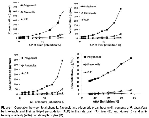
The total phenolic, flavonoids, and proanthocyanidins contents per each g dry extract of P. dactylifera L. were respectively 50.7 mg tannic acid, , 10.38 mg rutin, and 5.45 mg cyanidin. The polyphenolic, flavonoid, and proanthocyanin contents of extract showed an apparent linear relationship with the inhibition of lipid peroxidation of brain (r2 = 0.8466, p < 0.0001; r2 = 0.8322, p < 0.0001; r2 = 0.6707, p < 0.001), liver (r2 = 0.8070, p < 0.001; r2 = 7821, p < 0.001; r2 = 0.6349, p < 0.005 ), and kidney (r2 = 0.7296, p < 0.001; r2 = 0.7012, p < 0.001; r2 = 0.5558, p < 0.01 ), and erythrocyte hemolysis (r2 = 0.9848, p < 0.0001; r2 = 0.9524, p < 0.0001; r2 = 0.8448, p < 0.0001) ().
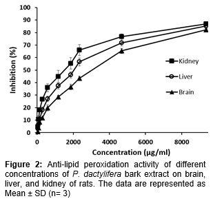
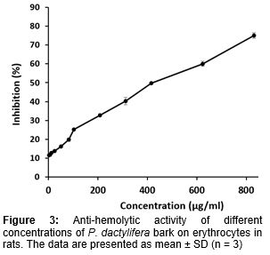
The IC50 for inhibition of lipid peroxidation in brain, liver, and kidney were 3150.72, 1941.45, and 1546.09 µg/mL, respectively (). The results showed that the bark extract effect on the kidney was higher than other tissues. Different concentrations of P. dactylifera bark extract were used to evaluate its effects on erythrocyte hemolysis in rat (3-850 µg/mL) (). The IC50 for P. dactylifera was 472.41 µg/mL. The results of correlation assessments between the main variables of the study are summarized in .
Discussion
Free radicals are continuously produced in the body via enzymatic and non-enzymatic reactions like respiratory chain reaction and phagocytosis. They damage biomolecules and polyunsaturated fatty acids are highly sensitive to oxidative stress. Lipid peroxidation is a free radical-related process in biologic systems that thiooxidants to modify the susceptibility of organs or tissues to oxidative stress and to alter the cellular antioxidant defense system in vitro.
The levels and types of polyphenol compounds have been widely studied for their antioxidant properties. Several mechanisms have been proposed for the polyphenol compound actions of prevention of oxidative stress and ROS/RNS generation both in vitro and in vivo but radical scavenging is the most widely accepted mechanism for their antioxidant activity [11]. Polyphenols in plants have several groups, such as flavonoids, proanthocyanidins, coumarins, and for most of them, antioxidant effects have been reported. According to the Pearson's correlation test, the order of the correlation between inhibition of lipid peroxidation of various tissues with polyphenolic groups was as follows: polyphenols > flavonoids > proanthocyanins. The mechanism of polyphenols in these assay can be direct radical scavenging and inhibition of lipid peroxide generation by chelating iron [12].
This study focuses on the malondialdehyde production as a product of lipid peroxidation. Several studies have demonstrated the relationship between malondialdehyde and pathophysiology of human disease [13].
Neuronal cells in the brain are highly sensitive to oxidative stress due to their dramatic dependence on oxidative phosphorylation for energy as compared to other cells and high level of fatty acids.
The demand for oxygen consumption is extremely high with 1 - 2 % of the oxygen being converted into superoxide anion radicals and hydrogen peroxide, leading to oxidative stress. The brain is most affected by lipid peroxidation because of its high oxidizable lipid, high metal content and lower in antioxidant activity in comparison with other tissue [14,15].
Clinical evidence shows that neurodegeneration can be ameliorated upon dietary or supplementary intake of natural antioxidants. Dietary intake contains variety of antioxidants and vitamin supplements which play a vital role in neuro-protection in variety of neurological disorders [14,15]. These natural antioxidants prevent oxidation of proteins, lipid peroxidations and prevent generation of ROS.
To evaluate the effect of P. dactylifera bark on the rat brain tissue, different concentrations of the extract were used. The IC50 value for P. dactylifera was 3150.72 µg/mL ().
Inhibitory effects of lipid peroxidation have been studied on different plants. Bagchi et al studied the inhibition of lipid peroxidation of grape seed`s proanthocyanidins on brain tissue and observed 50 % inhibition of lipid peroxidation in 100 mg/mL concentration [16].
In this study, good correlations were found between inhibition of lipid peroxidation of brain and polyphenol, flavonoids, and proanthocyanin. Cheung et al evaluated the anti-lipid peroxidation effect of mushroom (Lentinus edodes) and observed that the aqueous extract of L. edodes has a significant anti-oxidative activity against lipid peroxidation of rat brain with the IC50 value of 1.05 mg/mL [17].
Liver is among organisms that are attacked by lipid peroxidation because it contains high amounts of lipid. This causes several diseases such as aging, cancer, and liver fibrosis [18]. Different herbaceous compounds such as polyphenols and flavonoids are powerful lipid peroxidation scavengers in the liver [19]. Different concentrations of extract were used to evaluate the effect of P. dactylifera bark extract on the liver tissue of rat. The calculated IC50 for P. dactylifera was 1941.45 µg/mL. Sanchez et al studied the anti-lipid peroxidation effect of Mangifera indica on the liver and concluded that in concentrations of 50 and 250 mg/mL, oxidation inhibition is 46 and 52 %, respectively [20]. According to the Pearson's correlation test, there is good correlation between polyphenol, flavonoids, and proanthocyanin and inhibition of lipid peroxidation in the liver.
Srinavasan et al studied the anti-lipid peroxidation effect of Caesalpinia digyna root on kidney and observed that the plant has high scavenging power and appropriate therapeutic potential [21]. Kang et al studied the protective effect of sesame oil against lipid peroxidation and observed that the rats received diet containing sesame oil extract for 14 days, have a kidney with a high protective power against lipid peroxidation [22]. Results of this study showed good correlations between inhibition of lipid peroxidation of kidney and polyphenol, flavonoids and proanthocyanin.
The red blood cell is substantially susceptible to oxidative stress due to the high content of polyunsaturated fatty acids in the membrane and the auto-oxidation of haemoglobin within the cell. In addition, RBCs are always exposed to high concentration of oxygen and should be under anti-oxidative protection [23].
The IC50 for P. dactylifera inhibition of rat erythrocyte hemolysis was calculated as 472.41 µg/mL. Lekse et al studied the anti-lipid peroxidation and antioxidative effects of catechol extracted from some plants. For 5 mg/mL concentration of catechol, they observed a protective effect against lipid peroxidation in the membrane of red blood cell [24]. Mickstacka et al studied the anti-lipid peroxidation effect of RBC by quercetin and concluded that quercetin with IC50 of 64 ± 8.7 µM is powerful in protecting RBC [25].
Despite difference between ability of the extract on inhibiting of lipid peroxidation in various tissues, there is a significant correlation between assays. This can be attributed to the similarity in lipid tissues and oxidizing agent or the same mechanism of the effect of extract in the aforementioned assays.
Conclusion
The results of this study indicate that the bark of P. dactylifera is rich in phenolic compounds with antioxidant properties which can inhibit the lipid peroxidation and oxidative effect of free radicals. The antioxidant activity is stronger than on brain and liver tissues. Therefore, they can be used to treat diseases related to lipid peroxidation.
Declarations
Acknowledgement
References
Archives


News Updates